Cryogenic electron microscopy (Cryo-EM)
Since its introduction in the late 1970s, cryogenic electron microscopy (cryo-EM) has evolved from a low-resolution, so-called "blobology" technique to an established high-resolution structural biology method on its own.
This advanced technology combines cutting-edge methods in the preparation of frozen-hydrated samples, transmission electronic microscopy and image analysis, thus allowing the resolution of macro-molecular structures isolated in their near-native state or in situ:
- cryo-SPA (Single Particle Averaging) is a highly versatile structural biology technology that allows for the characterization, down to a few Å resolution, of isolated proteins of various sizes (<100 kDa to several MDa), origins (including membrane proteins) and quaternary states
- cryo-ET (Electron Tomography) opens the door to structural biology in situ, i.e. the structural characterization of biological macromolecules within their cellular environment, with resolutions better than 1 nm
The IECB cryo-EM facility proposes a complete workflow for the characterization of isolated proteins by low- and high-resolution SPA (negative stain and vitrified samples, respectively), as well as an integrated cryo-CLEM workflow (Correlative Light and Electron Microscopy) coupled to cryo-ET that allows for the characterizaton of fluorescently labelled macro-molecules in situ.
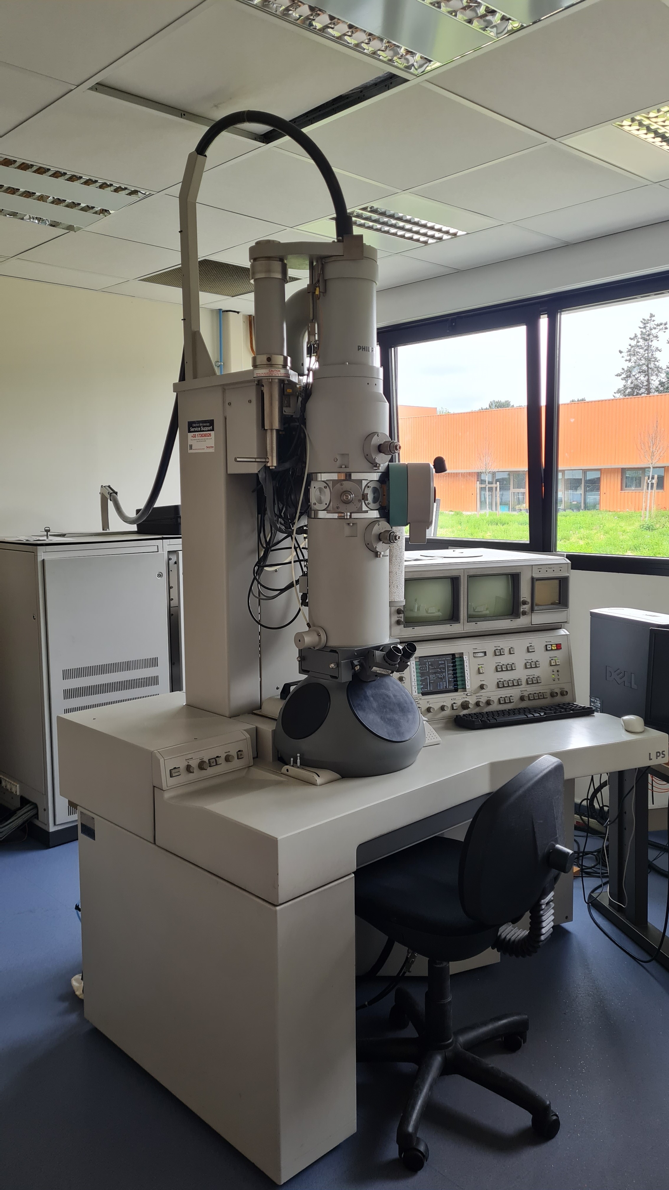
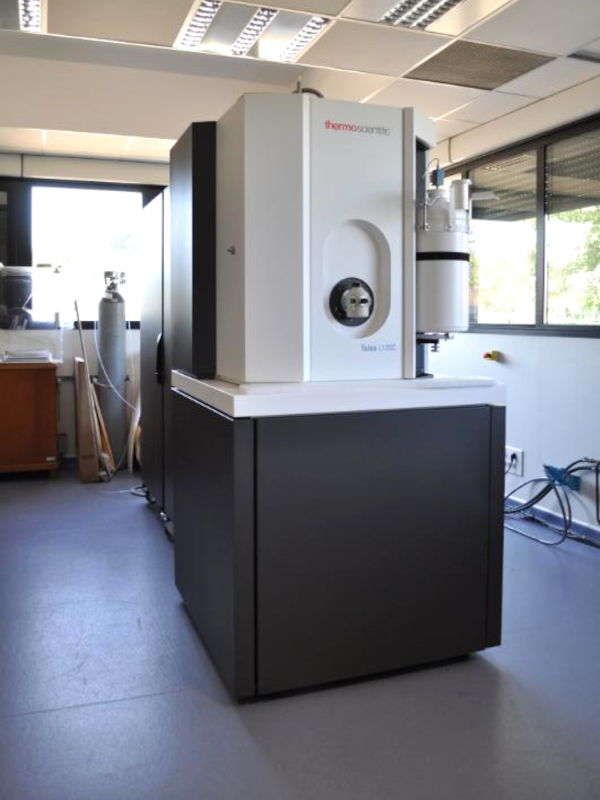
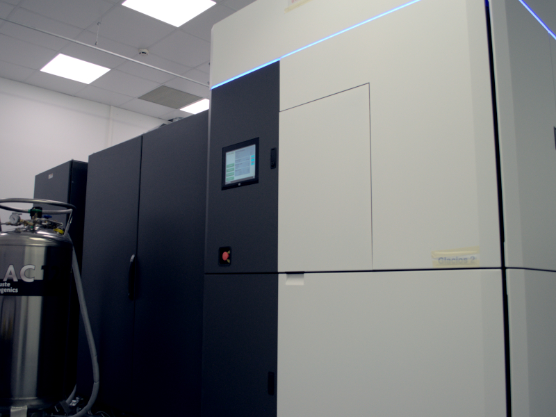
The facility is equipped with 4 electronic transmission microscopes, ranging from 120 kV to 200 kV, that allow for the characterization of polymers, nanoparticles, vesicles and biological macro-molecules.
Access:
For any analytical experiment with assistance, or for a R&D project, please ask the facility engineers for a detailed quotation:
- users can perform sample preparation and their analysis after training
- a full service can be provided by the research engineers
Facility Manager:
Axel Siroy : a.siroy@iecb.u-bordeaux.fr
Scientific Expert:
Rémi Fronzes : remi.fronzes@u-bordeaux.fr
Equipments
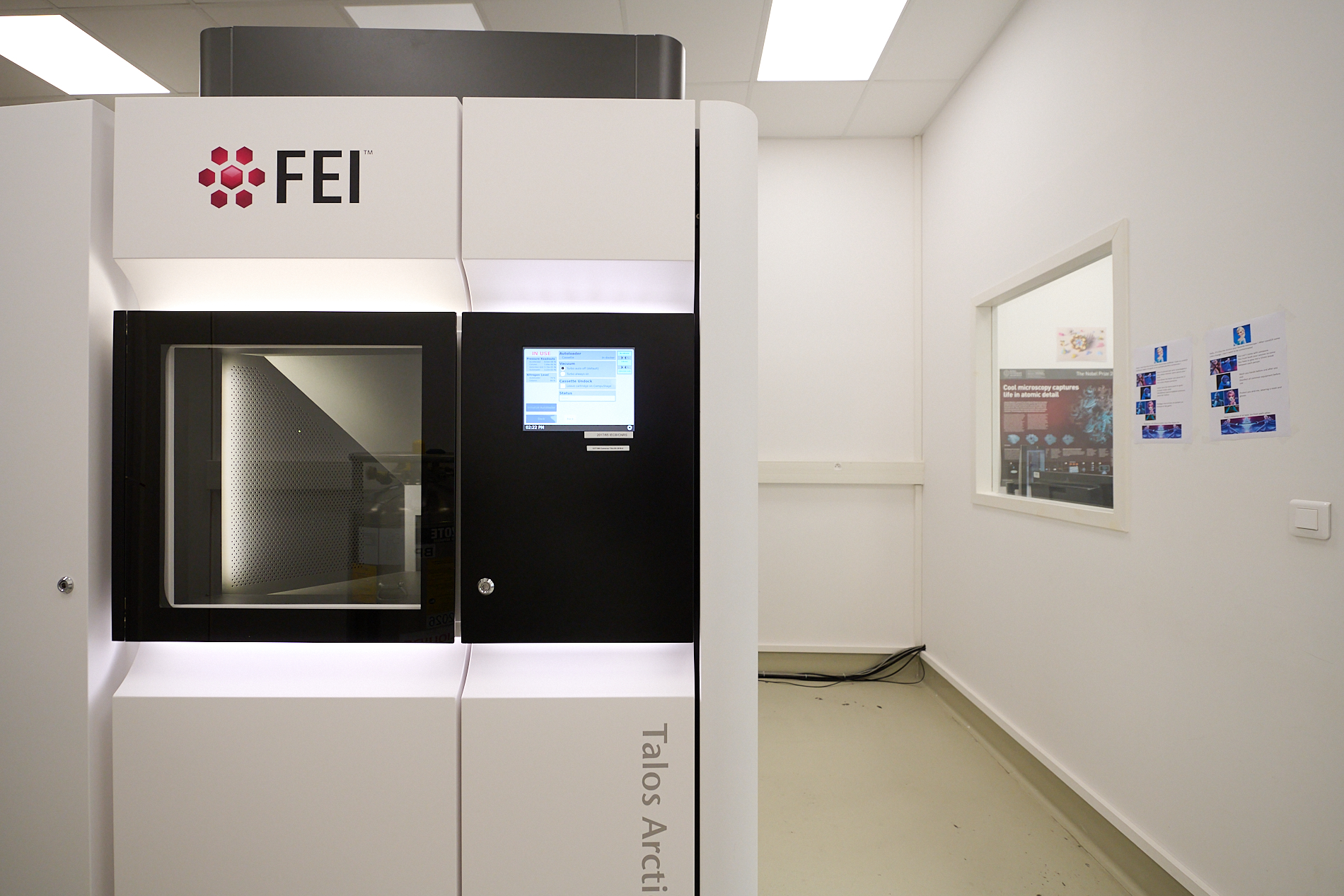
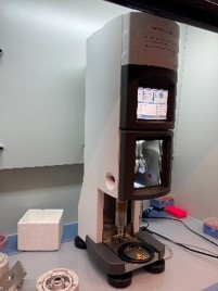
GLACIOS2 (FEI/ThermoFisher)
Latest-generation electronic transmission cryo-microscope
Automatic collection in cryo-SPA & cryo-ET
People
